Tag Archives: sample prep

magnifying glass

How Workflow Advances Raise the Bar in LC-MS/MS Veterinary Drug Quantitation

By Ed George
No Comments
magnifying glass

In the modern world, it’s often taken for granted that consumers can head to their local grocery store and fill their baskets with a broad range of meat, poultry, fish and dairy produce. Yet the plentiful availability of these products is possible, to a large extent, thanks to modern farming methods that rely on veterinary drugs to promote healthy animal growth, protect livestock from contracting diseases, and in some cases provide aesthetic qualities to food.

Despite the important role veterinary drugs play in farming and food production, usage must be carefully controlled, as their inappropriate administration can have adverse effects on animals, the environment and human health. A particular concern is the growing problem of antimicrobial resistance, which can be promoted in the environment by the overuse of some of these veterinary drugs.

As a result, the analysis of veterinary drugs forms an important part of routine food safety and quality control testing. However, the wide range of residue concentrations required to be quantified, along with the diverse sample matrices and chemical properties of multiple classes of veterinary drugs placed in a single analytical method, pose significant analytical challenges. The latest multi-residue, multi-class analytical workflow solutions using a generic sample preparation method and liquid chromatography tandem mass spectrometry (LC-MS/MS) are overcoming these issues to provide a robust, sensitive method for the extraction, detection, confirmation, and quantitation of veterinary drugs below their required maximum residue limits (MRLs).

Meeting the Needs of Veterinary Drug Analysis Workflows

Given the need to accurately and reliably quantify veterinary drugs in food, testing workflows must be both sensitive and operationally robust. Importantly, workflows must be amenable to a variety of different matrices, including meat, fish and dairy, and should be capable of screening for drug molecules with a broad range of physicochemical properties. The sample preparation protocols that are employed must minimize the loss of analytes and be sufficiently simple and cost effective to enable routine laboratory use. Additionally, the separation steps that are employed must be sufficiently rugged and should ideally be able to handle any analyte and matrix. Finally, the methods used to identify and quantify samples must be sufficiently selective and sensitive to detect and confirm drug molecules and their metabolites at trace levels.

Developing methods that can meet all of these criteria for a wide range of drug molecules and food matrices, while minimizing the potential for false positive and negative results, is not straightforward and has proven challenging for the industry. As a result, many analytical methodologies have emerged that are typically limited in scope to a limited number of residues or specific chemical classes, are labor intensive, and require extensive sample preparation and clean-up. Fortunately, ongoing advances in veterinary drug analysis workflows are helping to drive the adoption of standardized protocols that have universal applicability.

QuEChERS: Making Sample Preparation Quick, Easy and Reliable

Sample preparation is a key first step in veterinary drug analysis workflows, but its importance is often overlooked. Even with the most advanced downstream separation and detection technologies, workflows are liable to generate poor quantitative results without reliable residue extraction methods.Having robust sample preparation protocols is especially important given the heterogeneous nature of the sample matrix and the different physicochemical properties of the residues that must be extracted.

Traditional approaches, based on sample homogenization and multi-step solvent extraction procedures, were time-consuming and did not always produce consistent results. The loss of residues during sample grinding or through the formation of insoluble drug-matrix complexes would often impact the accuracy of measurements. Moreover, the need for labor-intensive sample cleanup steps, based on separation methods such as gel permeation chromatography, added additional complexity to workflows.

The widespread adoption of universal sample preparation protocols based on QuEChERS (Quick, Easy, Cheap, Effective, Rugged and Safe) methods has simplified the process of extracting veterinary drugs from matrix samples. These approaches have been specifically designed to be quick and easy to implement, and enable high extraction efficiency with a very broad range of chemical properties from a variety of matrices. As a result, QuEChERS has proven to be a very reliable means of preparing samples for veterinary drug analysis.

The universal suitability of the QuEChERS approach has reduced the complexity of sample preparation workflows to such an extent that many suppliers now offer kits containing pre-weighed reagents that can be used straight from the box. Moreover, because they only require small amounts of sample and solvent, and little in the way of equipment, these easy-to-use methods are helping laboratories minimize waste and make workflows more cost-effective.

Triple Quadrupole MS: Design Improvements Driving Exceptional Sensitivity

LC-MS/MS has rapidly established itself as the go-to technique for sensitive and reliable veterinary drug analysis, with set-ups based on ultra-high performance liquid chromatography (UHPLC) systems and triple quadrupole mass spectrometers proving to be particularly effective. With drug residues typically on the parts per billion scale, these systems have proven to be more than capable of delivering the level of performance that’s required when working with analytes that require low detection limits.

What’s more, recent advances in triple quadrupole mass spectrometer technologies are pushing the limits of quantitation even further. Improved instrument designs based on segmented quadrupoles, more powerful electron multipliers and enhanced ion transmission optics are enabling food analysis laboratories to achieve even greater levels of experimental sensitivity, mass accuracy, selectivity and precision. These performance improvements are allowing analysts to make more confident decisions around every sample.

ion chromatogram, salmon extract sample
Figure 1. Total extracted ion chromatogram of salmon extract sample at 1× STC. Results obtained using a Thermo Scientific Vanquish Flex Binary UHPLC system and a Thermo Scientific TSQ Altis triple quadrupole mass spectrometer. (Click to enlarge)

The capabilities of the latest generation of triple quadrupole LC-MS/MS systems for quantitative veterinary drug analysis were put to the test in a recent study. More than 170 veterinary drugs were added directly to a variety of homogenized matrices, including bovine muscle, milk, and salmon fillet using a QuEChERS sample preparation protocol to create a series of matrix extracted spikes (MES). The concentration of residues in the MES samples referenced a chosen screening target concentration (STC), which was typically one-third to one-quarter of the defined European Union MRL for each residue/matrix combination. Figure 1 presents the total extracted ion chromatogram for an MES sample of salmon fillet at the STC, obtained with a binary UHPLC system and a triple quadrupole mass spectrometer.

For each analyte, calibration curves were constructed using replicate measurements of each of the MES samples at seven concentrations ranging from one-fifth to five times that of the STC. Figure 2 highlights the calibration curve constructed for ethyl violet, a therapeutic dye used in aquaculture, in

Calibration curve, salmon extract
Figure 2. Calibration curve generated for ethyl violet in salmon extract (0.2–5.0 ng/g). (Click to enlarge)

the range 0.2 to 5.0 ng/g (STC = 1 ng/g). The calculated method detection limit of 0.03 ng/g for this compound in salmon fillet demonstrates confidence in the results well below the minimum required performance limit (MRPL).

LC-MS/MS: Leading the Way in Workflow Robustness

With potentially hundreds of samples to analyze every week, veterinary drug analysis workflows not only demand the highest levels of sensitivity, but also exceptional speed and robustness.
One way in which greater throughput can be achieved is by using shorter instrument dwell times, an experimental optimization that allows more compounds to be analyzed within a given timeframe during a chromatographic separation. Traditionally, the use of shorter dwell times would typically require sacrificing some measurement sensitivity. However, the latest advances in triple quadrupole instrument design are ensuring short dwell times no longer come at the expense of analytical performance.

Timed selected dreaction monitoring (SRM) is an effective strategy that allows analysts to overcome this challenge to achieve sensitivity with high throughput. Using timed SRM, data acquisition occurs within a short retention time window. This reduces the number of transitions that are monitored in parallel for each residue peak, while ensuring consistent quantitation even at low concentrations. Instrument control system software can automatically optimize the SRM conditions across the chromatographic run, maximizing operational efficiency with minimal need for manual input.

Instrument uptime is another factor that is of paramount importance for veterinary analysis workflows. With large workloads and tight turnaround times, regular instrument recalibration and frequent maintenance can be a major frustration for busy food testing laboratories. UHPLC is renowned for its operational robustness and suitability for fast-paced routine screening workflows, and the latest instruments are taking this reputation to an even higher level.
Comparison of injections of bovine muscle extract
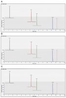
Figure 3. Comparison of injections of bovine muscle extract MES at 3× STC over 500 injections (A: injection #20; B: injection #260; C: injection #500). Analytes shown: cyromazine (black), ciprofloxacin (red), sulfamethoxazole (green) and flunixin (blue). (Click to enlarge)

Figure 3 compares injections of bovine muscle extract at 3× STC over a 500-injection run that took place over a period of one week, obtained using the experimental set-up described earlier. Despite continuous operation over this extended period, the peak shape, intensity and retention time stability are maintained. These results further highlight the robustness of the LC-MS/MS system for routine veterinary drug testing.

Conclusion

Enforcing the responsible use of veterinary drugs in farming and food production depends upon comprehensive, sensitive, robust and reliable workflows capable of delivering quantitative results. Advances in sample preparation techniques and LC-MS/MS technologies are setting new standards when it comes to confident multi-residue veterinary drug analysis. From the development of reliable easy-to-use QuEChERS protocols, through to robust UHPLC separation methods and sensitive triple quadrupole mass spectrometers, improvements across the workflow are driving exceptional performance—whatever the matrix, whatever the residue.

Debadeep Bhattacharyya, Thermo Fisher Scientific
In the Food Lab

Pushing The Limits Of Targeted Pesticide Residue Quantitation: Part 2

By Debadeep Bhattacharyya, Ph.D.
No Comments
Debadeep Bhattacharyya, Thermo Fisher Scientific

Detection and quantitation of pesticide residues in food is extremely important in food safety. Given the challenge of evaluating multiple pesticides at low levels across various samples, laboratories are in constant need of robust, reliable and sensitive analytical methods.

The risk of unauthorized pesticide overuse can increase residue concentrations in food, thereby, causing severe health issues. Global food safety bodies strictly regulate the levels of pesticides allowed in food products. In the European Union for instance, legislation in the form of Directive No 752/2014 sets statutory maximum residue limits (MRLs) for more than 1000 pesticides in food products of plant or animal origin.1 The number of pesticides and their allowed concentrations are necessary to ensure consumer safety, and are amongst the strictest in the world, permitting concentrations in products at levels typically as low as several parts-per-billion (ppb).

The requirements to achieve such low limits of quantitation for all analytes in a complex matrix present a significant analytical challenge for the food safety laboratories tasked with making a confident assessment of every sample. With perishable products such as fresh fruits and vegetables under routine analysis, these results need to be achieved within very short turnaround times and at a low cost per sample to meet lab managers’ budgets.

Advances in LC-MS/MS Technology

Recent advances in triple quadrupole technology have offered an additional boost to the existing analytical capabilities of liquid chromatography tandem mass spectrometry (LC-MS/MS). The segmented quadrupoles, faster rod drivers and more powerful electron multipliers can enable analysts to achieve the desired levels of robustness, mass accuracy, precision and sensitivity required to meet this challenge.

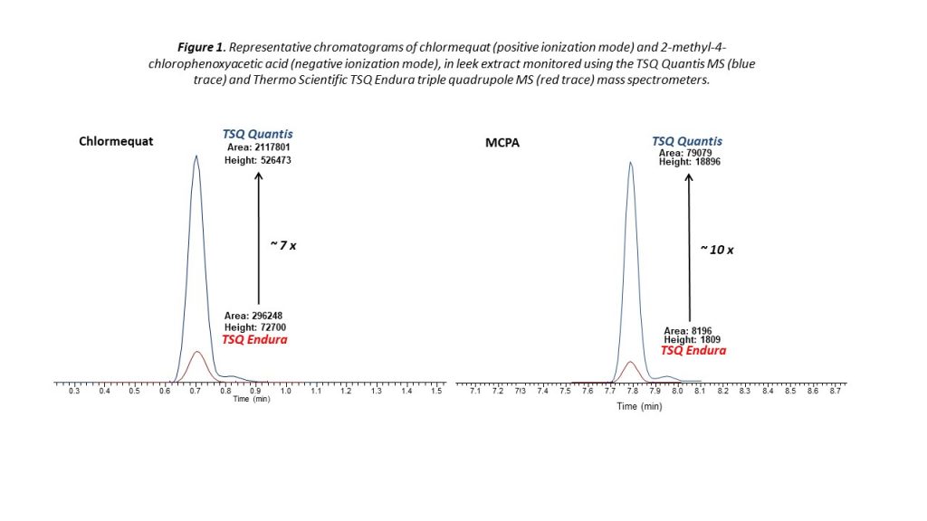
Improvements in instrument detection capability are pushing the limits of quantitation even further. Figure 1 highlights the amplified sensitivity of a triple quadrupole spectrometer for the determination of two pesticides in a leek sample—a particularly complex matrix with a high moisture content. For both chlormequat and 2-methyl-4-chlorophenoxyacetic acid (MCPA), the spectrometer delivers enhanced performance, giving analysts the ability to quantify residues far beyond the current limits required for MRL determination.

mass spectrometers
Figure 1. Representative chromatograms of chlormequat (positive ionization mode) and 2-methyl-4-chlorophenoxyacetic acid (negative ionization mode), in leek extract monitored using the TSQ Quantis MS (blue trace) and the TSQ Endura triple quadrupole MS (red trace) mass spectrometers.

Robust, Reliable and Reproducible

With potentially hundreds of perishable samples to analyze each day, food testing laboratories not only require the ultimate sensitivity, but sensitivity should be supported by speed and robustness.

One way in which analysts are achieving higher analytical throughput is through the use of shorter instrument dwell times. Although short dwell times in the past enabled productivity of sample analysis (more samples at the same time), they often came at the expense of robustness and sensitivity of the results. With the latest advances in triple quadrupole technology, short dwell times no longer compromise analysis.

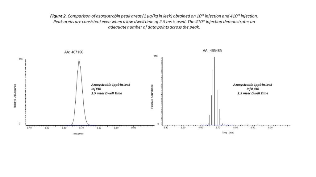
Very effective quantitation of pesticide residues can be achieved using timed selection reaction monitoring (SRM). With the timed SRM approach, data acquisition is performed within a short retention time window around each compound of interest. This approach reduces the number of transitions that are monitored in parallel within the retention time window, while ensuring consistent quantitation even at low concentrations (see Figure 2).

Pesticide Residue Quantitation
Figure 2. Comparison of azoxystrobin peak areas (1 μg/kg in leek) obtained on 10th injection and 410th injection. Peak areas are consistent even when a low dwell time of 2.5 ms is used. The 410th injection demonstrates an adequate number of data points across the peak.

Another important point to consider is workflow robustness. For busy laboratories with large workloads and tight turnaround times, time-consuming daily instrument recalibration and frequent maintenance simply isn’t a viable option.

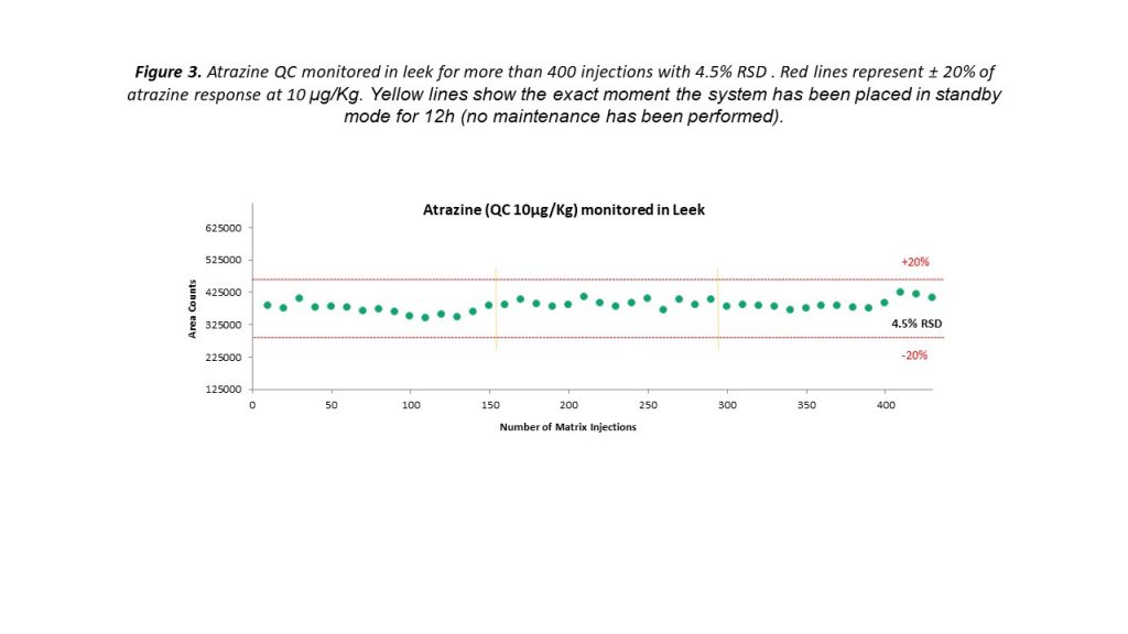
Triple quadrupole instruments are renowned for their experimental reliability that is delivered for every fast-paced environment, and the latest instruments are pushing expectations even further. Figure 3 demonstrates the precise levels of measurement reproducibility that can be achieved using a triple quadrupole MS. Peak areas for the pesticide residue atrazine, added to a leek sample at a concentration of 10 μg/kg, remained well within the expected ±20% range over 400 injections. Even when the system was placed into standby mode for 12 hours and subsequently restarted, consistent measurements could be obtained without any additional maintenance.

Pesticide Residue Quantitation
Figure 3. Atrazine peak areas (10 μg/kg in leek) monitored over 400 injections. Red lines represent ±20% atrazine response. Yellow lines show the point at which the system was placed in standby mode for 12 h. No system maintenance was performed between injections.

Conclusion

Technical advances in instrumentation and improvements in procedures have generated more robust LC-MS/MS processes to definitively detect trace pesticide residues. With limits of quantitation growing increasingly stringent year on year, such advances in technology are not only helping laboratories meet the quantitation challenges of today, but also prepare for those of tomorrow.

References

1. Commission Regulation (EU) No 752/2014 of 24 June 2014 replacing Annex I to Regulation (EC) No 396/2005 of the European Parliament and of the Council, 2014.

Acknowledgements

This article is based on research by Katerina Bousova, Michal Godula, Claudia Martins, Charles Yang, Ed Georg, Neloni Wijeratne & Richard J. Fussell Thermo Fisher Scientific, Dreieich, Germany, Thermo Fisher Scientific, California, USA, Thermo Fisher Scientific, Hemel Hempstead, UK.

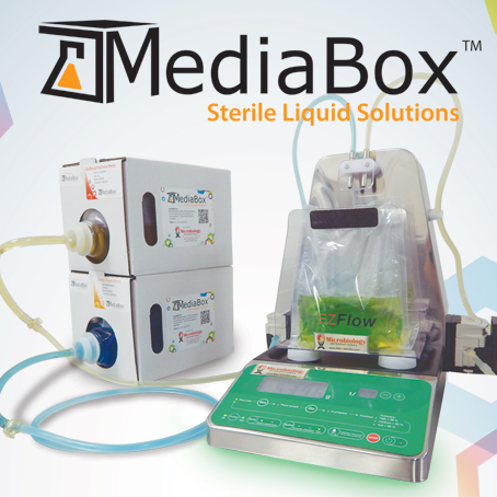
MediaBox Sterile Liquids, EZ-Flow Gravimetric Diluter Automate Sample Prep

MediaBox broths and buffers are sterile, easy-to-use and come in a convenient stackable storage box with a long shelf life. Significantly reduce staff workload by removing weighing, measuring, mixing, autoclaving and cleaning glassware. MediaBox is supplied ready-to-use and is far easier to use than dry bags, which are difficult to fill, often leak and are not consistent from one bag to the next.

MediaBox directly connects to the EZ-Flow gravimetric diluter creating an automated system for weighing and diluting your samples. EZ-Flow automatically weighs samples and provides diluent from MediaBox for the correct dilution factor. Your lab will love the convenience and increased efficiency. Microbiology International offers a wide range of dosing systems to pair with your MediaBox of choice.

All MediaBox products pass strict quality control protocols and include Certificate of Analysis documentation. MediaBox sterile liquids come in 5L, 10L and 20L boxes.

Available types include Buffered Peptone Water, Modified UVM, mTSB, Demi-Fraser Broth Base, Phosphate Buffer, Butterfields, Lactose Broth, Sterile Water, LB Broth, PBS, and more. Custom formulations are available.

Find the Weak Link in Pesticide Residue Analysis

By Food Safety Tech Staff
No Comments

Plagued by false or inaccurate results during pesticide analysis? To get to the bottom of the issue, it’s important to find any weaknesses in your pesticide residue workflow. In a recent blog on Analyte Guru, Richard Fussell of Thermo Scientific discusses areas in which pesticide testing labs can identify the weak links, including:

  • Solvent extraction. QuEChERs (Quick, Easy, Cheap, Effective, Rugged and Safe) acetonitrile extraction method
  • Sample processing
  • Extraction efficiency

Get the details about how to find the weakest link in Fussell’s blog here.

More on pesticide analysis:

Rapid and Robust Technologies Improve Sample Preparation for Analyzing Mycotoxins

By Olga I. Shimelis
No Comments

Mycotoxins are produced as secondary metabolites by various mold species during the growth and harvest of grains, fruits, nuts and condiments. Their production is directly related to the dry/wet weather conditions during the growing season. Mycotoxins are very stable compounds and are not easily removed during storage, processing and preparation of raw agricultural commodities.

Mycotoxins & Grains
Mycotoxins can be found in a variety of grains.

Different classes of mycotoxins are distinguished on the basis of the structural similarity and originating mold species. For example, more than a dozen different aflatoxin compounds exist but only five of them are routinely tested (aflatoxins B1, B2, G1, G2, and M1). Aflatoxin B1 is of particular interest because it is listed as a Group 1 Carcinogen by the International Agency for Research on Cancer (IARC). Aflatoxin M1 is a metabolic product that can be present in milk upon ingestion of aflatoxin B1 by an animal. Aflatoxins are ubiquitous in important agricultural commodities including maize and peanuts, and are among the most studied mycotoxins.

Deoxynivalenol (DON) is produced by a different fungi species. It is prevalent in cereal crops grown under wet conditions and temperatures above 15o C (60o F). Chronic exposure of livestock to DON may result in slowed growth, impaired immune function and reduced rates of reproduction, particularly in non-ruminants.

Mycotoxins were discovered as the cause of poisoning outbreaks in both humans and farm animals in the mid-20th century. Since then, multiple government regulations were established to control the presence of these toxic compounds in food and feeds. For example, harvested grains are checked for mycotoxin contamination using rapid field screening methods prior to grain deposition into silos. If contamination is found, the crops are sent to an analytical laboratory to perform the confirmation analysis. Liquid chromatographic methods were often used for such analysis with both fluorescence and UV detection. In recent years, mass spectrometry has been employed as a detection method.

Sample Preparation for Laboratory Mycotoxin Analysis

When performing analysis, it is important to choose the right sample preparation method to ensure accuracy, sensitivity of detection, repeatability and robustness, as well as fast sample preparation for high throughput. During laboratory analysis of mycotoxins, the sample preparation procedure typically includes extraction, purification and concentration steps.

Extraction of mycotoxins from samples is conducted by mixing the ground sample with the mixture of organic solvent and water, such as acetonitrile:water (80:20). Using methanol is not recommended, because it does not provide complete extraction. Prior to cleanup, the sample is filtered. Historically, mycotoxin analysis required extensive extract cleanup to minimize interference by matrix components. This holds true as new regulations continue to require lower detection limits.

Cleanup methodologies often include the use of phase extraction (SPE). Of the different types of SPE, one of the most common is the use of immunoaffinity sorbents that result in the selective retention and cleanup of mycotoxins. The drawback to using the immunoaffinity sorbents in the lab is that they are not compatible with the mycotoxin extraction solvent. In order to load the extract into the immunoaffinity SPE tube, the extract must be diluted with water, sometimes 20-fold, to prevent precipitation or folding of the protein-based antibodies by exposure to organic solvent. This presents an additional sample preparation challenge, as the grain extracts tend to form precipitates upon the addition of water and can clog the SPE columns. Thus, apart from the high cost of immunoaffinity SPE columns, the methods tend to be labor and timeintensive.

Super Tox SPE cartridges
Super Tox is a line of SPE cartridges for mycotoxin families that eliminates extra sample prep steps.

It would be beneficial to a laboratory to eliminate these extra sample preparation steps required by immunoaffinity SPE. Such cleanup SPE procedures are available and can be applied directly to the mycotoxin extracts without the need for further dilution, filtration and evaporation. A line of SPE cartridges for different mycotoxin families was recently introduced to the market. These SPE cartridges are compatible with the extracts generated during mycotoxin extractions and can be stored at room temperature. The tubes can also be used for cleanup of multiple classes of mycotoxins.

Analysis of Aflatoxins and Zearalenone

SPE cartridges are available for aflatoxins and zearalenone.

The following results employed SPE cartridges for mycotoxins that can be used for two aflatoxin classes, aflatoxins and zearalenone, and were applied to the cleanup of grain and peanut extracts. Results were compared to cleanup using immunoaffinity columns.

AflaZea SPE cartridges are based on the “interference removal” strategy that requires fewer processing steps compared to the “bind-and-elute” strategy of the other SPE. Peanut extracts contain not only co-extracted protein and complex carbohydrates but also fat. This extract was successfully cleaned using AflaZea SPE. When the SPE tube and a leading IAC column were applied to the peanut extract, both methods demonstrated good recoveries for spiked aflatoxins B1, B2, G1, G2 with AflaZea recovery values of 101–108% and immunoaffinity recovery values of 79–100%. However, the AflaZea provided better reproducibility for detection with a relative standard deviation (RSD) of 2–4% RSD versus 10–25% RSD with immunoaffinity SPE. This is likely because sample preparation using AflaZea is less tedious and takes one tenth of the time compared to immunoaffinity SPE.

Analysis of Deoxynivalenol

Wheat samples can be analyzed for deoxynivalenol using a new SPE cartridge.

The following compares a new SPE cartridge for the analysis of DON, one of the Fusarium mycotoxins, with immunoaffinity SPE. Analysis of DON often is conducted using liquid chromatography (LC) with UV detection, so sample cleanliness is important to permit the separation of the DON peak from background interferences. The new SPE DON cartridge was compared to the immunoaffinity SPE for the cleanup and analysis of wheat samples. Clean chromatography and good recovery of spiked DON was obtained by both methods (86–97% RSD). However, clogging of the filters by the immunoaffinity SPE sample was observed during cleanup and complicated the sample preparation procedure. The SPE DON cartridge provided faster sample preparation.

Analysis of Patulin

Patulin is a mycotoxin commonly found in rotting apples.

Another SPE technology for mycotoxin analysis is based on molecularly imprinted polymers (MIPs), which are sometimes called “chemical antibodies” and mimic the performance of immunoaffinity sorbents. MIPs have binding sites that conform to the shape and functionality of specific compounds or compound classes. Strong binding of the analyte to the MIP makes it possible to perform intensive SPE washes that lead to very clean samples. Unlike immunoaffinity sorbents, MIPs are compatible with organic solvents and strong acids and bases.

Foods containing apples and similar fruits are required to be tested for patulin toxin, as they are the most common source for patulin exposure in humans. The MIP SPE procedure for patulin is faster than other SPE or liquid-liquid extraction methods and provides selective retention and superior cleanup. It is a robust method for analyzing apple juice and apple puree with HPLC-UV detection. After cleanup, patulin is quantified in apple puree at 10 ppb levels, which meet most regulatory requirements. The MIP SPE cleanup method eliminated 5-(hydroxymethyl)furfural (HMF) from the matrix, which sometimes appears as an interfering chromatographic peak when other sample prep methods are used. An SPE wash using sodium bicarbonate removed the interfering organic acids, while patulin was stabilized during elution at the end of the SPE procedure by using acidified solvent. Thus, most problems encountered during patulin analysis were resolved during this single SPE procedure.

Conclusion

As government regulations and consumer demand warrant cleaner, non-contaminated products, mycotoxin analysis will continue to be performed around the world. Careful selection of sample preparation methods is required for such analysis to achieve accurate testing results, best method performance and high laboratory throughput. Although many sample preparation methods exist, laboratories should choose the methods that not only provide adequately prepared samples, but also result in time and cost savings. The SPE technologies discussed in this article are sample preparation techniques that provide the required analytical sensitivity without capital expenditure into higher-end LC-MS equipment; the LC-UV and LC-FL methods can still be used. In addition, these SPE methods are simple, more robust, and less-time consuming compared to other SPE methods or liquid-liquid extraction.

All images courtesy of Sigma Aldrich

Aaron Kettle is the Product Manager, Thermo Fisher

Need a Faster Sample Prep Method for Pesticide Residue Analysis? Try Accelerated Solvent Extraction

By Aaron Kettle
No Comments
Aaron Kettle is the Product Manager, Thermo Fisher

In a Q&A with Food Safety Tech, Aaron Kettle, sample preparation product manager at Thermo Fisher Scientific discusses the advantages of using accelerated solvent extraction for pesticide residue analysis and its applications in the food industry.

Food Safety Tech: What are the benefits of using accelerated solvent extraction versus other more time-consuming sample prep methods?

Aaron Kettle: It can save a considerable amount of time over techniques such as Soxhlet and sonication that are used in this industry. In addition, the amount of solvent you’re using is cut down by at least five fold using this technique. The other advantage over some of the other techniques is that it allows walk-away capability, so your samples can be loaded and you can run your methods overnight. It’s pretty much just load the buttons and walk away, and by the time you come in the next day, everything is ready for analysis.

FST: Discuss this method in the context of today’s environment as it relates to importance of detecting the presence of pesticide residue and harmful pollutants.

Kettle:
It’s certainly beneficial. It doesn’t have matrix limitations so you can use it for a lot of different sample types: High-fat content samples, such as avocados, dry samples like bread and grain, and high-water content samples like tomatoes.

FST: Are there specific food applications that benefit from accelerated solvent extraction, including those in which the method is underutilized?

Kettle: I don’t think the technique has a lot of use right now for high-water content samples, such as pesticide residue extraction for tomatoes, berries, etc. That primarily has to do with the fact that historically it hasn’t worked well with these kinds of samples. However, we’ve recently released a moisture-absorbing polymer that acts to remove residual water without interfering with the extraction and recovery of the analytes. That has allowed the accelerated solvent extraction to work with these sample types. That’s an area where’s it’s being underutilized right now, primarily because it is a relatively new product release for us. It’s an area where we’d like to see adoption continue to increase.

FST: What is unique about the polymer being used in this detection method?

Kettle: It’s a proprietary mixture that will remove water content up to 85%. There are no major specifics; it will work with any kind of matrix that has water in it—fruits, berries, etc.

FST: What are the top advantages of the technology?

Kettle: The ability to remove residual water is important. It happens prior to the sample being loaded in the extraction cell. There is no limitation with high-water content samples. It mixes well with the dispersing agent, so not only can you add a dispersing agent to help solvent flow through the matrix better, you can also add the polymer to help it through water. It helps expand the capability of the accelerated solvent extraction in what it can do in the food market for pesticide extraction.

FST: What are your expections of this technology within this niche moving forward?

Kettle: We would like to see it expand and have greater awareness and acceptance for the accelerated solvent extraction in this particular area. Right now, folks are using a manual technique for these types of samples, so we’re hoping these customers will accept the walk-away automation and the flexibility that this technique will provide.

Developing an Effective Environmental Monitoring, Sampling and Testing Program

By Food Safety Tech Staff
1 Comment

As the food industry is moving toward a more preventive food safety strategy, environmental monitoring is playing an increasingly critical role in testing. Hazard analysis is shifting the focus from finished product testing to proactively testing the environment and the processing as critical control points to continuously monitor and reduce risk. Today many facilities are adding or strengthening their environmental monitoring programs to enhance their food safety risk reduction efforts.

In a recent webinar, Ann Draughon, Emeritus Professor of Food Microbiology and Toxicology, University of Tennessee spoke about Developing an Effective Environmental Monitoring, Sampling and Testing (EMS) Program. We present some excerpts from her presentation.

What do you need to get started with an EMS program?

“You need to first identify the right team; think about what kind of food you are processing (raw products or ready-to-eat products) and if it has had any food safety outbreak associated with it; determine critical or hygiene zones in your facility; determine sample locations; finalize which indicator tests will be done, and in which zones; determine which pathogens you will test for; choose the right test methods; set a baseline, and link that with your sampling plan, and establish testing frequency once you have finalized the number of samples and zones,” explains Draughon.

To establish critical hygiene zones, she advises to:

  • Survey entire facility and have a map of that facility;
  • Study that map and identify traffic patterns to divide the facility into critical hygiene zones, GMP zones, and non-processing zones;
  • Put in place barriers between these zones and dedicate equipment to the critical hygiene zone, and restrict access between zones; and
  • Establish strict cleaning, sanitation and monitoring plans for these diff zones.

Sampling of zones should be based on risk of contamination and/ or transmission of pathogens to food from environment, says Draughon. The sampling should also take into account potential sources of product contamination by whatever means during food processing (see image 1 for examples of 4 zone and 3-zone hygiene systems).

Selecting the right assays for your EMS program

There are many options, and it can be confusing to select the right assay for your needs. Draughon advises that companies need to look their monitoring needs and consider both indicator bacteria and pathogenic bacteria to select the right assay.

For monitoring with indicator bacteria, most companies look at ATP for environmental sanitation, often before start-up to make sure facility is clean before processing begins. Protein assays are also used to pick up any allergen on equipment.

APC or total viable count is a simple assay offering many choices, which tests for the number of live bacteria on your equipment or in your environment that can grow under air or oxygen at room temperature.

Yeast/ mold count assays are good for two purposes: 1. Mold frequently is the cause of spoilage in food, so it’s useful to understand if there are any present to determine shelf life, and 2. It also helps us understand the number of particulates in the air.

We can also select specific microbial groups as indicators, such as total Enterobacteriacae, fecal coliform or E.coli or Listeria species.

Sample collection and prep

When we collect a sample, we have to clearly document the sample including information such as when it was taken, from where, by whom, what happened to that sample etc. Use clean SOPs to reduce error. Use the assays previously selected and do it as quickly as feasible. If you are working with an outside company, decide how they are going to handle the sample. Finally, always keep in mind plant safety and leave nothing behind after sampling, and avoid cross-contamination.

For characterizing pathogens, you may want to genetically fingerprint any pathogenic isolates from your facilities. This will allow you to see if you have a constant harborage of a particular pathogen or if it changes. Draughon recommends using a contract lab for characterizing pathogens, as they would be better suited, and have better resources to do this. Destroy the isolates after characterization – you don’t want any chance of the pathogen spreading into the product or the environment.

Written SOPs for EMS programs

It’s critical to have clear written SOPs for EMS programs which include the following:

  • Frequency of sampling;
  • When, where , how and duration of sampling;
  • Procedure for recording data and coding;
  • Sample number, size or volume;
  • Specific sampling and analysis validated protocols;
  • Monitoring of incubators and use of equipment;
  • Handling and shipping of samples; and
  • Alert and action levels and appropriate response to deviations from alert or action levels.

It’s also important that we train and validate the personnel performing EMS. Each individual doing this needs to demonstrate proficiency of doing this. They need to understand proper recording of EMS program data, alert and action levels, and zero tolerance levels. The personnel should be comfortable and qualified for sampling protocol, and using all the equipment.

In summary, sampling plans should be adaptable, which highest risk sites being tested initially. Establish a baseline and modify sampling plan as needed. Establish your sampling and testing criteria and sample as needed with each zone to fully assess the environmental program.