Tag Archives: LC-MS

Jens Brockmeyer
Allergen Alley

HPLC-MS: Advancing Routine Food Allergen Testing

By Jens Brockmeyer, Eva-Maria Niehaus
No Comments
Jens Brockmeyer

In the U.S., allergens are the sixth leading cause of chronic illness with over 50 million people affected annually.[1] This is just one country among many that has seen the steady increase of the prevalence and severity of food allergies.[2]With increased media attention on this growing issue, food product labeling and allergen testing have never been under such scrutiny.

In food product labeling the stakes are high. Consumers need to know that the ingredient lists of the products they buy are accurate, and manufacturers need assurance that their foods are allergen-free, especially as current labeling guidelines lack clarity.

Food testing methods, which have hitherto been deficient in detecting and identifying unknown allergens and cross-contamination within the food manufacturing process, must be further developed to become quicker and more precise, cost effective, and user-friendly. New analytical tests can identify a broader range of potential allergens and offer food manufacturers a way to detect emerging allergens.

The Pressure for Improved Regulation

With the number of people suffering from food allergies in the U.S. doubling in each of the last two decades,[3] there is a high demand for food manufacturers to improve the allergen information they provide for consumers.

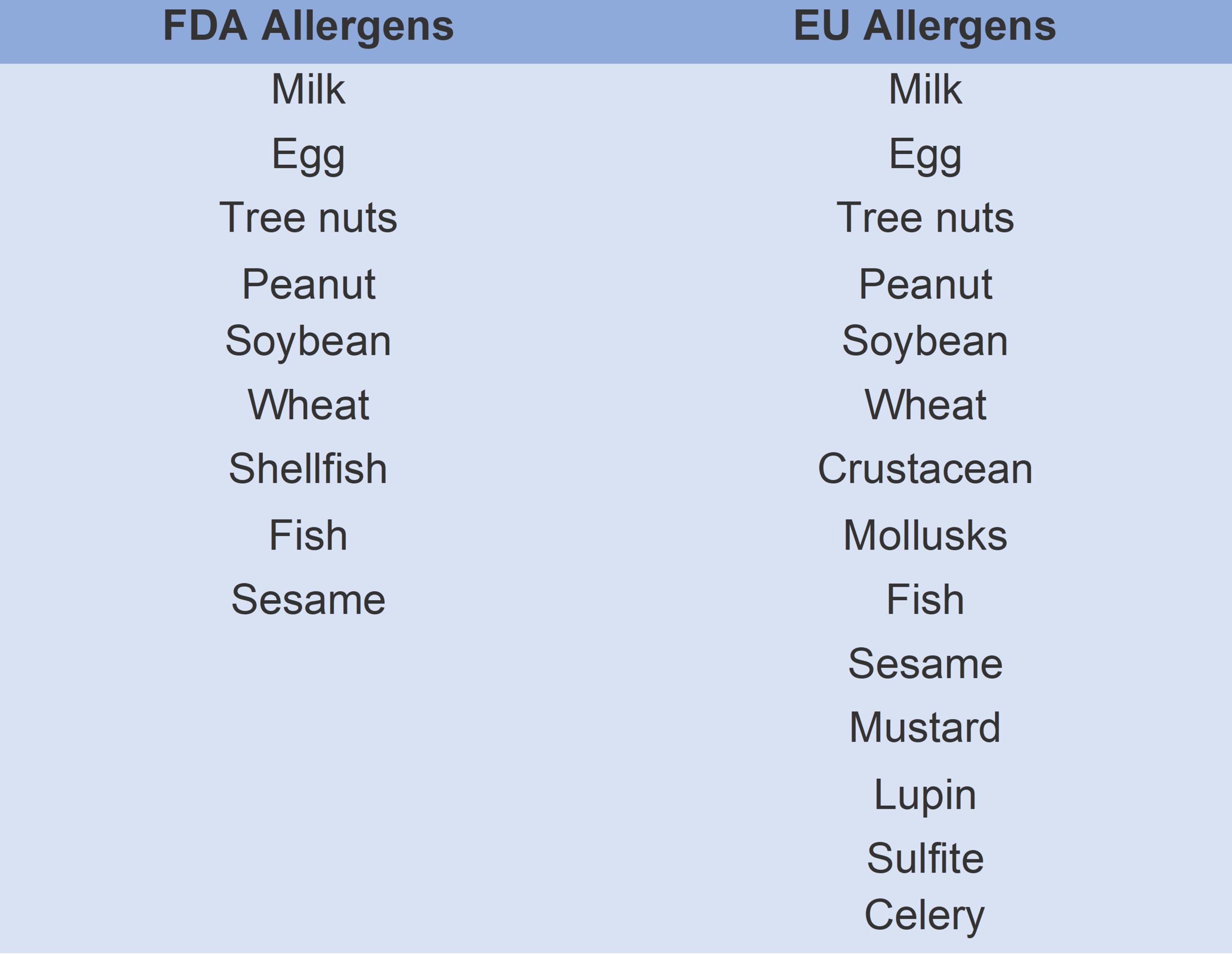
However, there is a lack of uniformity across different regions. While European Union law stipulates that allergens must be listed in bold on product ingredient lists, only 14 of the 200 foods that could potentially cause allergic reactions are prioritized[4] and, in the U.S., the FDA lists only nine key allergens.[5] Furthermore, while organizations such as Anaphylaxis UK agree that the most severe allergic reactions are caused by the consumption of a certain quantity of an allergen, there is no universal agreement on what such threshold levels should be.[6]

The allergens regulated by the FDA and EU.
Figure 1: The allergens regulated by the FDA and EU.

Single ingredients are not the only cause of allergic reactions: contamination and cross-contamination can occur at various stages of the food manufacturing process and put consumers at even greater risk.[7] Many manufacturers voluntarily use Precautionary Allergen Labeling (PAL) on their products to mitigate the risk of undeclared allergens, which may be used when there is a risk of allergen cross-contamination in the supply chain.[8] An example of PAL is ‘may contain milk’. This is not a legal requirement, however, and PAL protects the manufacturer more than the consumer as it is unlikely to be based on an assessment of the risk of cross-contamination for each of the 14 regulated allergens.[9]

Current Allergen Testing Options

Enzyme-linked immunosorbent assay (ELISA) and polymerase chain reaction (PCR) are the two main analytical methods presently used to detect and quantitate allergens in routine food testing.[10]

Highly sensitive, the ELISA method allows for the quantitative and qualitative analysis of antigens, proteins, hormones, and antibodies in biological samples. It is most commonly used in the identification of foodborne microorganisms, such as Salmonella and L. monocytogenes, across a range of food products.[11]

Although ELISA can identify specific analytical targets, it cannot detect unknown allergens in contaminated food supplies. Moreover, the overall structure of proteins and their extractability is often altered during food processing, which can affect assay test results. Other factors, including poor comparability of results between test kits that use different antibodies, can also impact each test, and therefore make reproducibility between methods difficult.

A comparison of ELISA, PCR and MS.
Figure 2: A comparison of ELISA, PCR and MS.

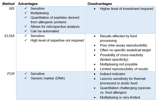
PCR testing, which uses deoxyribonucleic acid (DNA) as a genetic marker, is popular because of its sensitivity and specificity, and also because sample preparation is standardized. A limitation of PCR testing is that, as an indirect indicator, it lacks sensitivity for foods that could contain high quantities of allergenic protein, but little to no DNA.[12]

Liquid chromatography-mass spectrometry (LC-MS) is not commonly used in current food testing. Unlike ELISA, LC-MS has multiplexing capabilities, allowing for the detection of multiple allergens in a single run. It can provide precise separation, identification, and quantification of the specific peptides rather than proteins in samples, which not only increases test accuracy, but also improves upon traditional testing methods by allowing for the differentiation of closely related allergens.

However, LC-MS, too, has limitations. The complex matrices of some biological samples can cause issues, and the various sample techniques needed for specific analytes mean that implementing a LC-MS workflow in routine testing laboratories is difficult. Professor Jens Brockmeyer and his team at the University of Stuttgart have been working on further advancing LC-MS to mitigate such drawbacks.

The Solution for Comprehensive Allergen Testing

A research team at the University of Stuttgart is investigating the influence of food processing on allergenic potential. The team is seeking to improve the methods used to screen for allergens primarily through the use of mass spectrometry (MS) and has developed a new workflow for allergen testing that delivers results quickly and efficiently.

There are three components to the novel method: sample preparation, analysis by high-performance liquid chromatography (HPLC) coupled to MS, and data analysis software. The approach can be used in the analysis of food products and allergens, making sample preparation easier and simplifying laboratory workflows.

The workflow for the allergen screening method.
Figure 3: The workflow for the allergen screening method.

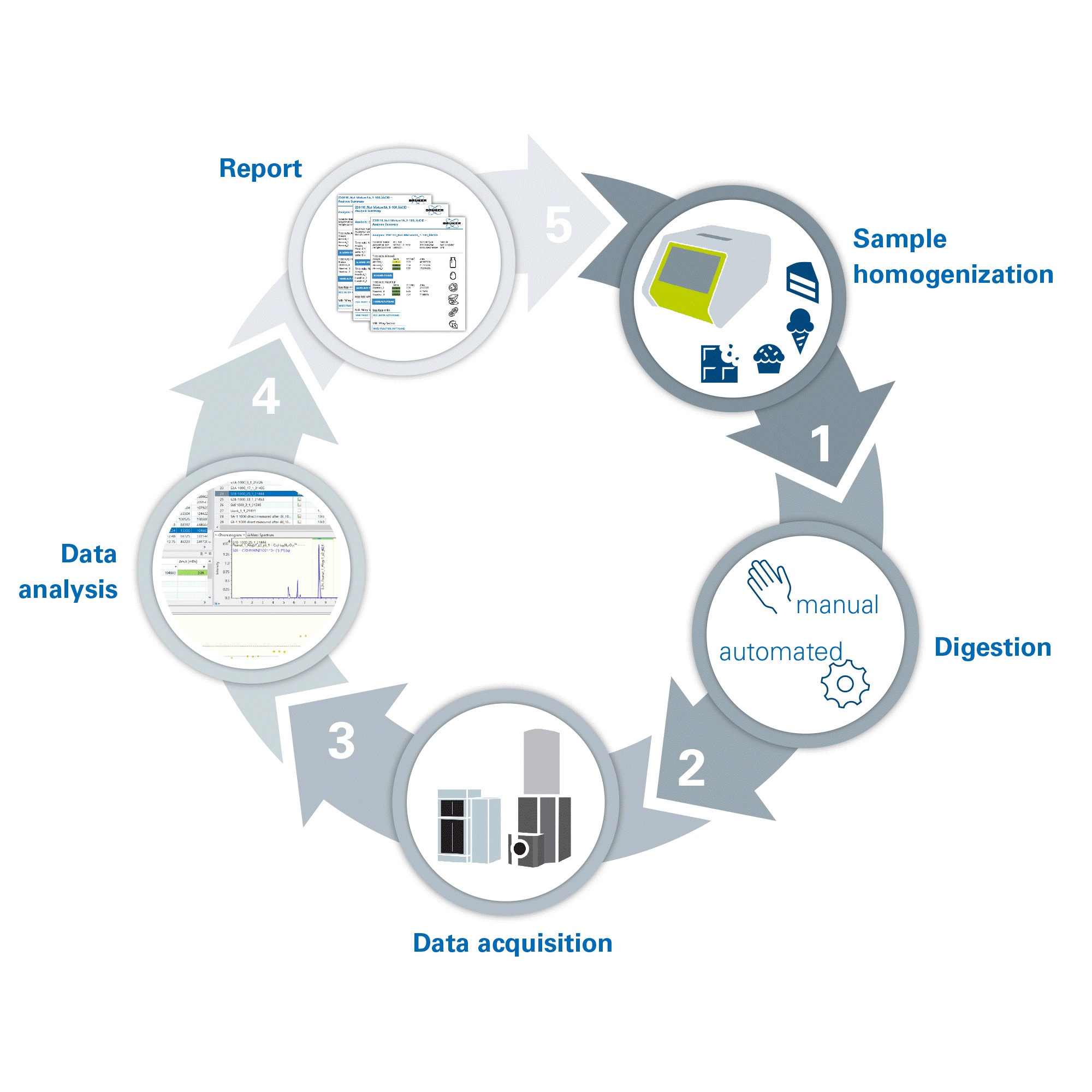
Protein extraction, manual or automated enzymatic digestion, and the cleanup of peptides takes place to prepare the sample. Protein digestion takes three hours, a considerable time saving in comparison to the usual proteomic procedures.[13] HPLC-MS is used to generate data; the detection of precursor masses of the specific peptides resulting from the allergenic proteins of the given ingredient verifies the presence of each allergenic element.

The new multiplexed method removes the need to run multiple tests to identify each allergen individually; just one run is required for the detection of several allergens. This multiplexing capability and the analytical software’s ability to evaluate measurements retrospectively significantly accelerates experiment time.

Improved Visibility of Unknown Allergens

Current food testing methods, such as ELISA and PCR, are valued for their sensitivity but both lack multiplexing capabilities and produce results that are affected by food processing and thermal processing, respectively. HPLC-MS is an innovative method that offers multiplexed analysis of complex samples, producing standardized results in an accelerated timeframe suited to the high throughput needs of food testing laboratories.

With undisclosed allergens the cause of 42% of food product recalls in the U.S. in 2022,[14] the precision and speed of HPLC-MS offers exciting potential for the future of food allergen testing, paving the way for the feasible implementation of clearer and more stringent regulations.

References

[1] American College of Allergy, Asthma, & Immunology. Allergy Facts. Accessed at: https://acaai.org/allergies/allergies-101/facts-stats/. Last accessed: November 17, 2023.

[2] The Guardian. ‘It’s one of the great mysteries of our time’: why extreme food allergies are on the rise – and what we can do about them. Accessed at: https://www.theguardian.com/society/2023/jul/15/its-one-of-the-great-mysteries-of-our-time-why-extreme-food-allergies-are-on-the-rise-and-what-we-can-do-about-them. Last accessed: November 17, 2023.

[3] Food Allergy & Anaphylaxis Connection Team. Food allergy & anaphylaxis. Accessed at: https://www.foodallergyawareness.org/food-allergy-and-anaphylaxis/prevention/food-allergies-on-the-rise/#:~:text=The%20number%20of%20people%20with,identified%20food%20allergy%20(source). Last accessed: December 21, 2023.

[4] European Union. Annex 2 – allergen labelling. Accessed at: https://food.ec.europa.eu/system/files/2018-07/codex_ccfl_cl-2018-24_ann-02.pdf. Last accessed: November 15, 2023.

[5] U.S. Food and Drug Administration. Allergic to sesame? food labels now must list sesame as an allergen. 2023. Accessed at: https://www.fda.gov/consumers/consumer-updates/allergic-sesame-food-labels-now-must-list-sesame-allergen. Last accessed: November 8, 2023.

[6] Anaphylaxis UK. Allergen thresholds. Accessed at: https://www.anaphylaxis.org.uk/fact-sheet/allergen-thresholds/. Last accessed: November 16, 2023.

[7] Food Allergy Canada. Avoiding cross-contamination. Accessed at: https://www.foodallergycanada.ca/living-with-allergies/day-to-day-management/avoiding-cross-contamination/. Last accessed: November 16, 2023.

[8] Food StandardsAgency. Precautionary allergen labelling. Accessed at: https://www.food.gov.uk/business-guidance/precautionary-allergen-labelling. Last accessed: November 16, 2023.

[9] Food Standards Agency. Precautionary allergen labelling. Accessed at: https://www.food.gov.uk/business-guidance/precautionary-allergen-labelling. Last accessed: November 16, 2023.

[10] Allergen Bureau. Food allergen analysis. Accessed at: https://allergenbureau.net/food-allergens/food-allergen-analysis/. Last accessed: November 16, 2023.

[11] Law JW-F, Ab Mutalib N-S, Chan K-G, Lee L-H. Rapid methods for the detection of foodborne bacterial pathogens: Principles, applications, advantages and limitations. Frontiers in Microbiology. 2015; 5.

[12] Stoyke M, Becker R, Brockmeyer J, et al. German government official methods board points the way forward: Launch of a new working group for mass spectrometry for protein analysis to detect food fraud and food allergens. Journal of AOAC International. 2019;102(5):1280-1285.

[13] Switzar L, Giera M, Niessen WM. Protein digestion: An overview of the available techniques and recent developments. Journal of Proteome Research. 2013;12(3):1067-1077.

[14] U.S. PIRG Education Fund. Food for thought part 2: An analysis of food recalls for 2022. Accessed at: https://pirg.org/edfund/resources/food-for-thought-an-analysis-of-food-recalls-for-2022/. Last Accessed: December 21, 2023.

Veterinary Drugs Analysis, Food Safety

Veterinary Drugs Analysis to Ensure Food Safety

By Olga I. Shimelis
No Comments
Veterinary Drugs Analysis, Food Safety

Monitoring for veterinary drug residues is conducted to ensure food safety and compliance with approved veterinary medicine practices. Veterinary drugs are used in animal husbandry for a variety of reasons, including as a curative/preventive of disease in herd and flock, to improve meat quality, and to promote growth. The chemical classes of drugs that may be used are broad, but major classes include antibiotics, anti-parasitics, and hormones. While risk-modifiers are used to minimize risk for consumption, residues from these drugs, their breakdown metabolites, or associated impurities of the drug may persist in animal tissue, necessitating the requirement that contaminant testing be undertaken.

In the United States, trace analysis of contaminants in food products began in the early 1970s following amendments to the Federal Food, Drug, and Cosmetic Act (FFDCA) in 1968. Worldwide, the regulatory requirements for contaminants in food have seen significant tightening due to a number of high-profile contamination crises and increased trade of food across country borders. From the technology standpoint, lower detection limits have been made possible by improvement of the detection capabilities of the analytical methods and instruments. Some of the most stringent requirements for contaminants in food are found in the European Union, where the levels of contamination should be below Minimum Residue Limits (MRLs), whereas in the United States, such limits are called U.S. tolerances.

Veterinary Drugs Analysis, Food Safety
Image courtesy of MilliporeSigma

When analyzing for drug residues, the choice of tissue has historically been the liver and kidney tissues, as these organs serve to remove the contaminants from the body and, as a result, the concentration of contaminants there is higher and easier to detect. Muscle tissue now often is added to the target list, as its contamination would have a direct impact on consumers.

With regards to veterinary drugs testing, one can distinguish between screening methods and confirmatory methods. The former should be fast and high-throughput and used to detect the presence of an analyte. The confirmatory methods should be able to provide confirmation of an analyte’s identity and quantitation at the levels of interest. Microbiological methods were popular for screening of antimicrobial drugs since these drugs inhibit growth of microorganisms, but suffer from a lack of specificity since not all microorganisms are equally sensitive to all antibiotics. Rapid screening methods include immunoassay-based testing kits, which are specific, fast, and can include multiple antibiotic classes in one test. Confirmatory methods typically include chemical analysis techniques with LC-MS detection, which provides the best ionization for most classes of veterinary drugs, along with better selectivity for focused analysis and lower detection limits. LC-MS can provide specific analysis of compounds from multiple classes in the same run through either targeted MS/MS or non-targeted analysis of unknowns through high mass resolution methods. The speed of LC-MS analysis has improved with the introduction of ultra-high pressure liquid chromatography-MS (UHPLC-MS) instruments. In the last few years, UHPLC-MS methods simultaneously serve as screening and confirmation methods for multiple classes, so called “multi-residue methods”. Some of these methods use MS/MS detectors and some use high-resolution mass spectrometers utilizing time-of-flight and ion trap detectors. These methods now can provide fast turn-around time and better accuracy in comparison to microbiological methods. They may be preferentially used by testing laboratories that are equipped and capable of utilizing the latest MS instrument technologies.

The 4th Annual Food Labs conference provides practical solutions and best practices on running, managing and equipping a food lab. | March 7–8, 2016, Atlanta, GA | LEARN MOREAll mass spectrometry methods that strive to perform simultaneous analysis of multiple veterinary drug classes are prone to the same drawbacks. Due to the differences in the analytes’ polarity, acidity and hydrophobicity, the quantitative extraction of analytes from tissue samples could be difficult. Ideally, the sample preparation methods should be compatible for compounds with varying physico-chemical properties but still provide selective separation from the matrix components to avoid occurrence of matrix effects during quantitation. The co-extracted matrix impurities are undesirable since they can affect the ionization of targeted analytes and result in under- or over-estimation of their concentration (ion suppression or enhancement). Due to the difficulty in designing a method that works for a wide variety of analytes, cleanup is often omitted for multi-class multi-analytes methods, and the stable isotope internal standards are used to correct for ionization effects during quantitation. However, omitting the sample cleanup could lead to other methodology problems.

As noted in the veterinary drug analysis session during the 2015 AOAC Annual meeting, sample cleanliness can result not only in matrix effects and impact quantitation, but it can also have an effect on the mass accuracy when high-resolution mass spectrometry is used and, therefore, can affect the identification of the analytes and lead to false negatives.

The most often used methodologies for sample cleanup during analysis of veterinary drugs in tissues is solid-phase extraction (SPE), both in cartridge and dispersive formats. C18 SPE proved to be a very versatile sorbent that often resulted in the best cleanup and best precision of analysis, closely followed by polymeric sorbents when applied to multi-class LC-MS analysis.

Aminoglycosides Antibiotics

Aminoglycosides is one class of veterinary antibiotics that is hard to include into multi-class methods. The aminoglycoside structures include connected modified sugars with different number of substituents including hydroxy- and amino-groups. The higher degree of polarity for aminoglycosides contributes to their solubility properties: these compounds are freely soluble in water and to some extent are soluble in lower alcohols, but are not soluble in common organic solvents and have solubility issues in solvent-water mixtures with high organic contents. Therefore, the normal extraction conditions that include organic solvents and are frequently applied to most other classes of veterinary drugs do not work well for aminoglycosides. A separate method is often used to extract and analyze these antibiotics.

Most often aminoglycosides are detected by mass spectrometry through the formation of positive ions during electrospray ionization. The LC separation of aminoglycosides could be done by either a reversed-phase (RP) method with ion-pair mobile phase additive to insure the retention of compounds or by HILIC chromatography. We have investigated both methods and looked at the sensitivity for detection of these compounds. The use of ion-pair is most often presented as a disadvantage, as it can reduce the analyte signal through the decrease of ionization efficiency and fouling the LC-MS instrument. While the use of ion-pair in our study decreased the ionization for some of the lighter compounds in this class (streptomycin, puromycin), ionization efficiency increased for the heavier mass compounds (gentamycin, neomycin). RP chromatography resulted in improved separation of the analytes compared to HILIC. LC-MS fouling from the use of HFBA was not observed in our investigation that spanned the course of a couple of years. In the HILIC mode with use of formic acid as a mobile phase additive, the detection of neomycin was problematic due to very low sensitivity. It was as low as one seventh of the sensitivity obtained by RP method.

The instrument response for aminoglycosides also depends on sample extraction and cleanup and the accompanying matrix ionization effects. The extraction from animal tissues has been traditionally done using the McIlvaine buffer that includes 2% Tricloroacetic acid (TCA) to precipitate proteins and release any bound analytes and 0.4 mM EDTA to prevent the binding of the analytes to cations and/or glass. Then the extract undergoes cleanup steps using SPE. The SPE sorbent most often used is a cation exchange phase, as the aminoglycosides have ionizable amino-groups and can be retained from the extract through ion-exchange interactions. Another option for the SPE cleanup became recently available—molecularly imprinted polymeric (MIP) SPE. MIPs, which are sometimes called “chemical antibodies”, mimic the performance of immunoaffinity sorbents. MIPs have binding sites that conform to the shape and functionality of  a specific compound or a compound class. Strong binding of the analyte to the MIP makes it possible to perform intensive SPE washes that lead to very clean samples. Unlike immunoaffinity sorbents, MIPs are compatible with organic solvents and strong acids and bases.

Selective interactionWe have tested the MIP SPE versus the traditional weak cation exchange (WCX) SPE cleanup for aminoglycosides spiked into pork tissue. The resulting ionization effects were compared as an indication of samples cleanliness. The quantitation in both cases was done using matrix-matched calibration curves and in both cases the recoveries for most of the ten tested aminoglycosides were above 70% (with exception of spectinomycin at 33% in case of WCX cleanup and tobramycin at 55% in case of MIP cleanup). For the two cleanup methods, there was a significant difference in matrix effects. In Figure 1, matrix factors close to 1.0 indicate little to no matrix influence for analyte detection: the ionization of the analyte in mass spectrometer is not influenced by co-extracted matrix impurities and quantitation values are not skewed. Values for matrix factors that are significantly greater than 1.0 suggest matrix enhancement for the analyte and values less than 1.0 are considered to be the result of matrix suppression. Significant matrix suppression was observed for all analytes when WCX SPE was used for cleanup. The ion suppression effect was significantly less for samples cleaned using MIP SPE. In addition, we observed significant time savings when using the MIP SPE cleanup method, as it did not require sample evaporation after using water-containing elution solvent.

Figure 1. Matrix factors close to 1.0 indicate little to no matrix influence for analyte detection

Conclusions

While improvement in the laboratory instrumentation allows the simultaneous and fast analysis of multiple contaminants, sample preparation remains important for reliable identification of contaminants in screening methods and error-free quantitation in confirmatory methods. Both the extraction and sample cleanup methods can contribute to accurate multi-class methods analyzing wide variety of veterinary drugs. New and upcoming technologies such as molecularly-imprinted polymers could be used for more targeted analysis of specific classes of analytes via instrumental methods.